
Analog
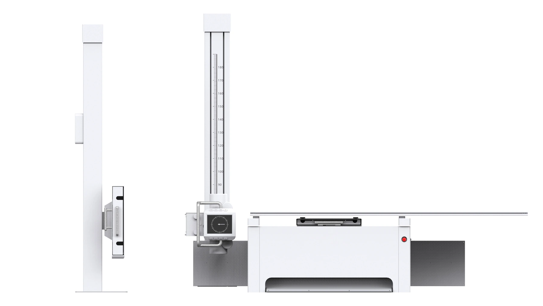
YJ-FDR100U-50
Floor-mounted analog X-ray system with a 50 kW generator, 40-150 kV range, 10-630 mA and 0.6 / 1.2 mm focal spots.
View model →The YJ-FDR200-50 combines a 50 kW X-ray generator, rotating-anode tube, 43 x 43 cm CsI flat panel detector, motorized tube stand, 4-way floating patient table and DICOM acquisition workstation. The system is configured for hospitals, clinics and imaging centers that need reliable table and upright radiography.
The values below describe the supplied 50 kW configuration. Generator output, detector type, wireless detector, grid, tube option and room layout must be confirmed against the final quotation and destination-market requirements.
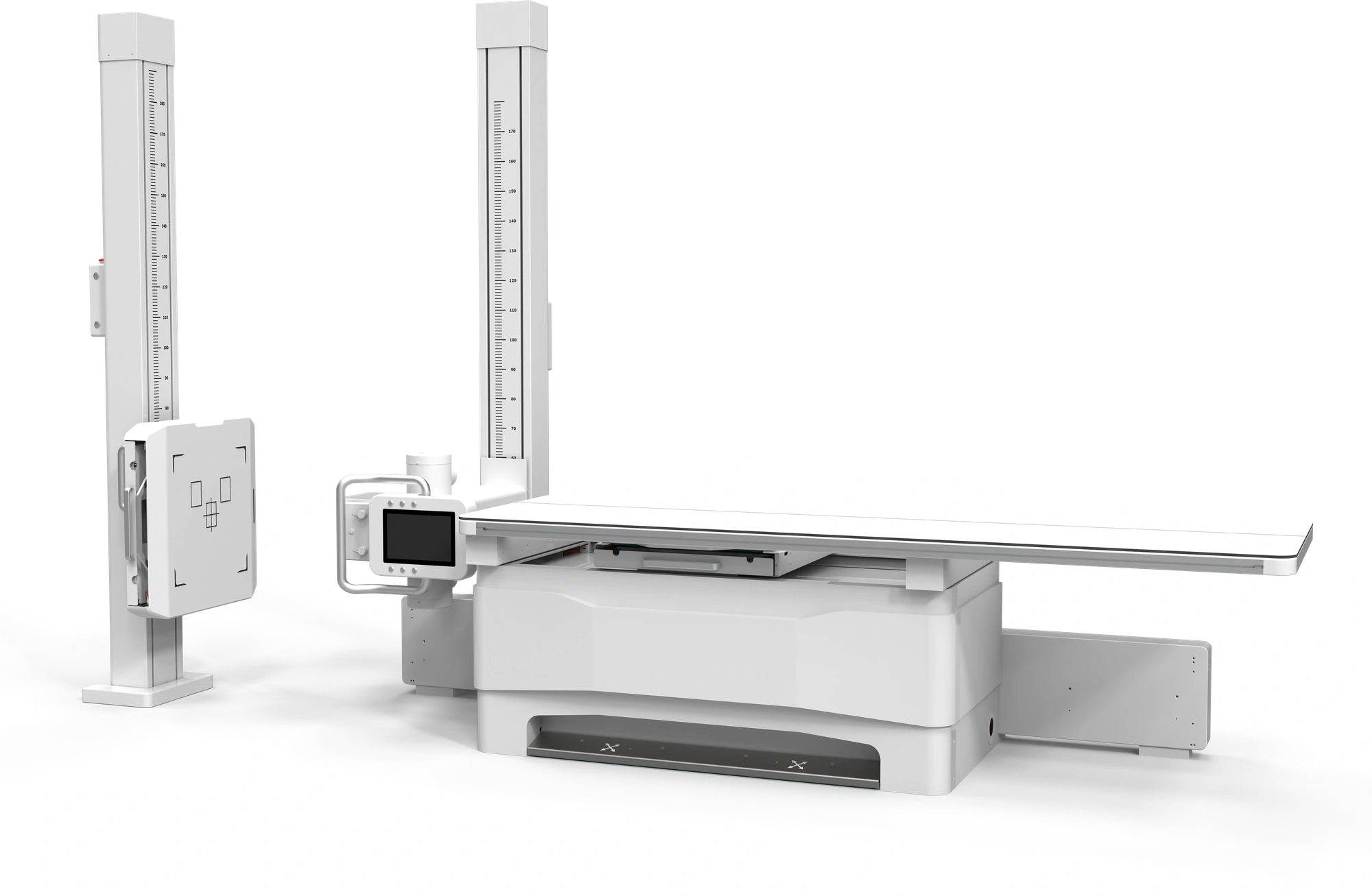
Start from the room workflow, generator output and detector configuration, then open a model page for the full specification. More fixed DR models can be added to this directory as they are released.
Floor-mounted analog X-ray system with a 50 kW generator, 40-150 kV range, 10-630 mA and 0.6 / 1.2 mm focal spots.
View model →50 kW floor-mounted DR with a 43 x 43 cm CsI detector, motorized tube stand, 4-way floating table and DICOM acquisition workstation.
View model →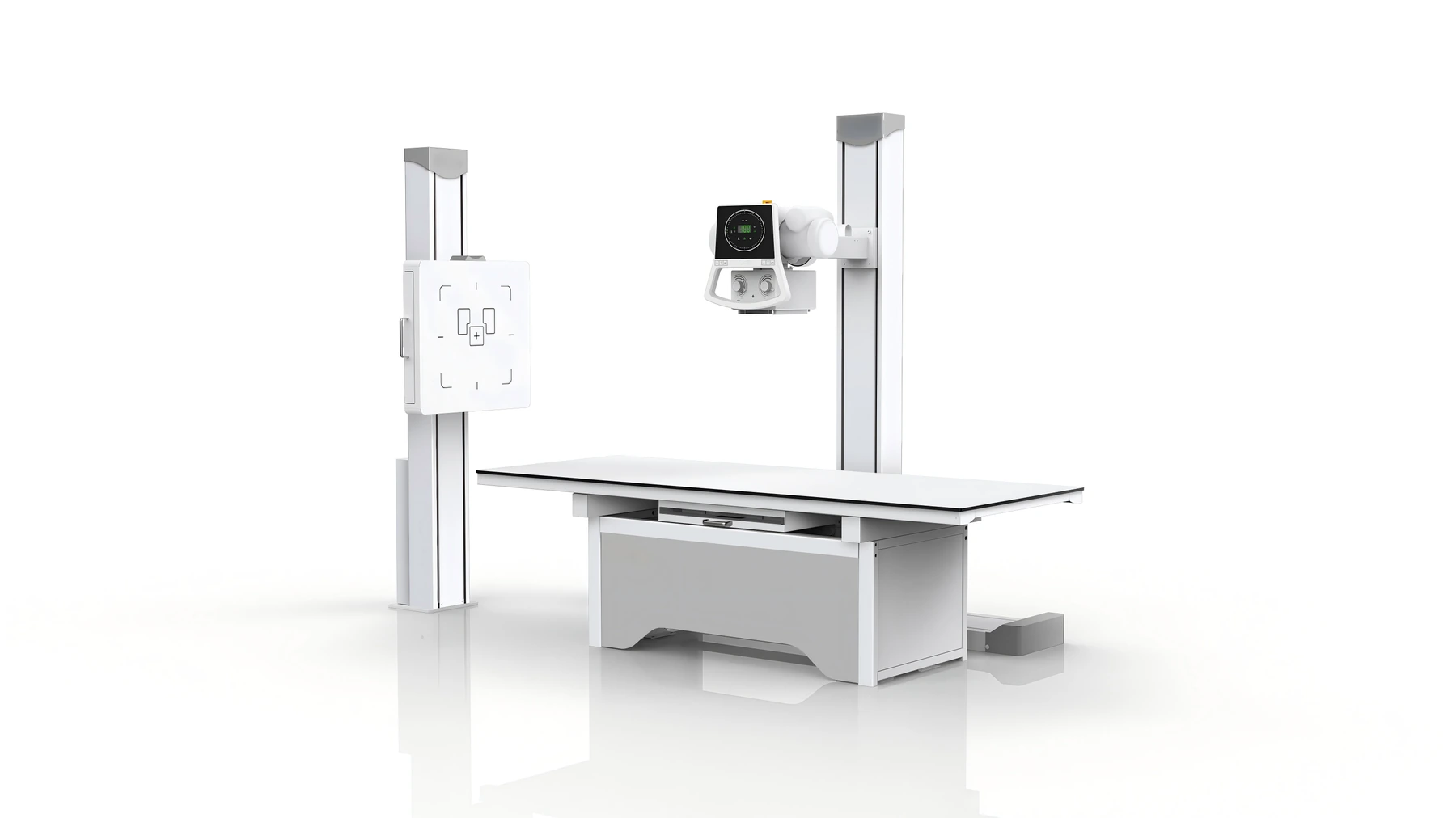
50 kW floor-mounted DR with a 17 x 17 inch CsI detector, 3072 x 3072 matrix, 139 um pixel pitch and a 24-inch workstation.
View model →The white-background views show the complete system layout. The installation image shows the same product in a real room setting.
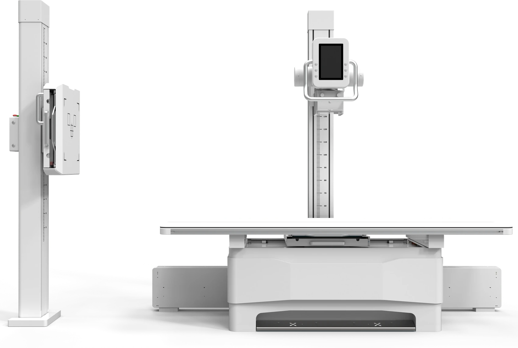
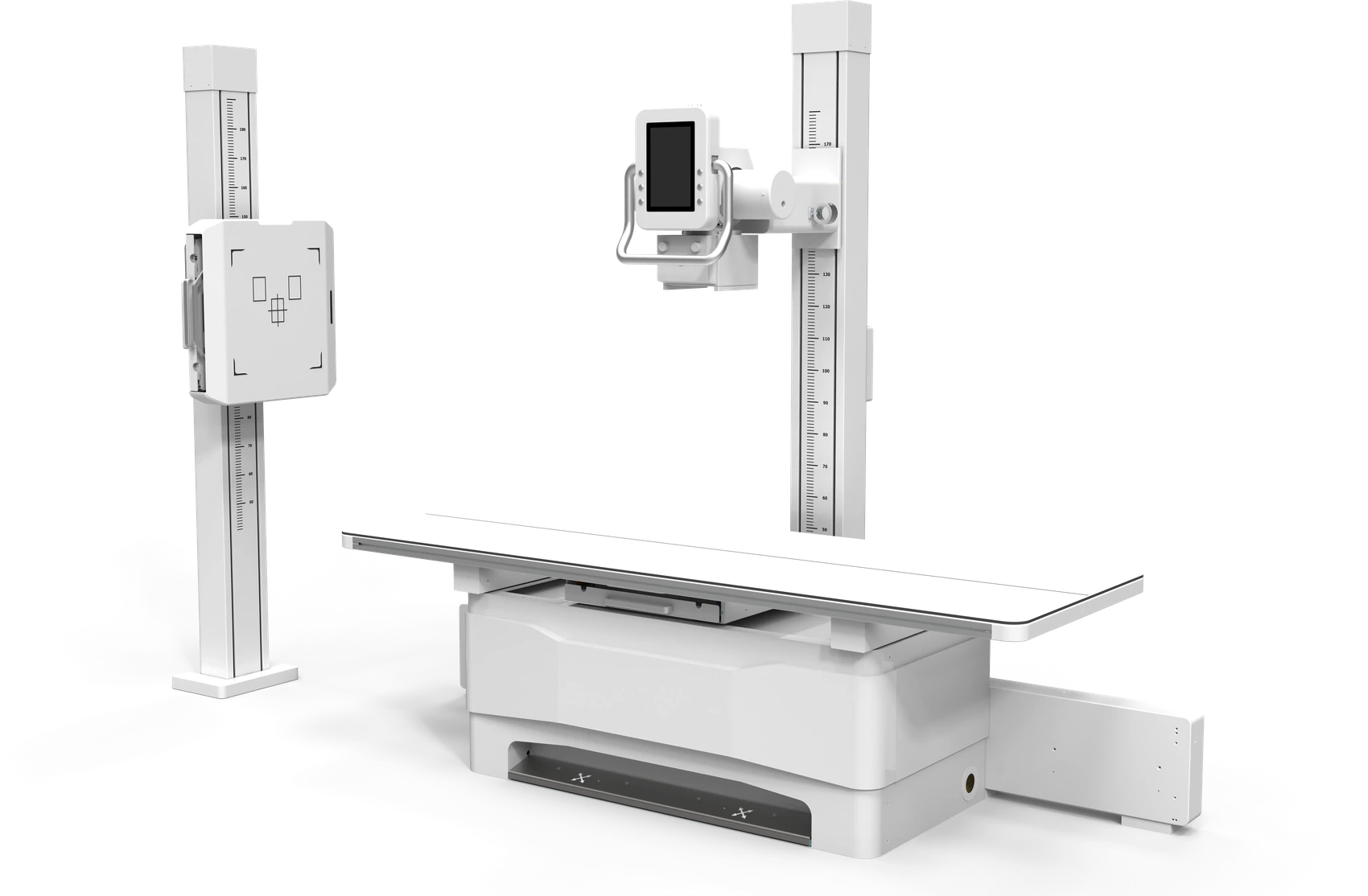
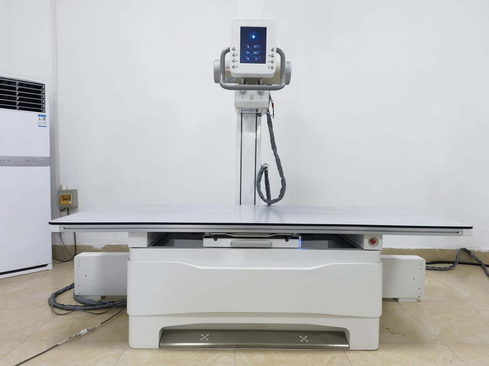
The supplied configuration focuses on positioning speed, clear operation and stable digital image acquisition.
Motorized synchronization between the tube stand and detector column supports faster table and upright positioning.
A high-resolution control screen keeps examination parameters and positioning controls close to the operator.
Supports standing knee anterior-posterior examinations for orthopedics, emergency and routine imaging workflows.
The 2200 x 800 mm tabletop provides front/back and left/right travel to reduce patient repositioning.
These photographs show the generator, tube, detector, control interface and workstation included in the product set.
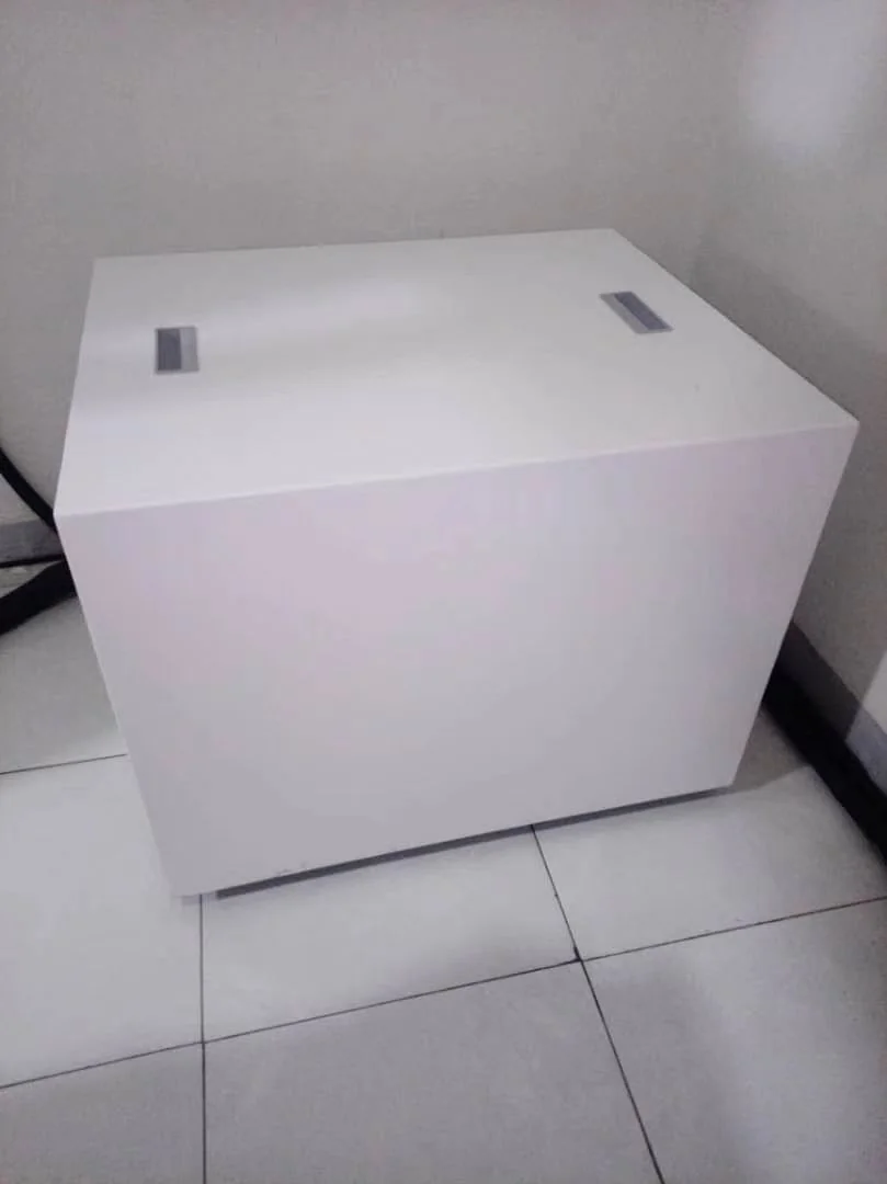
Self-protection and fault-code review support stable operation and service communication.
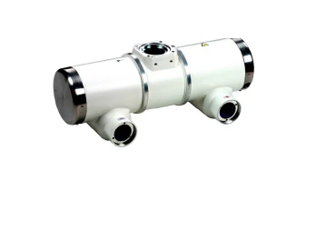
Dual 0.6/1.2 focus, 300 kHU anode heat storage and 750 W maximum heat dissipation in the 50 kW configuration.
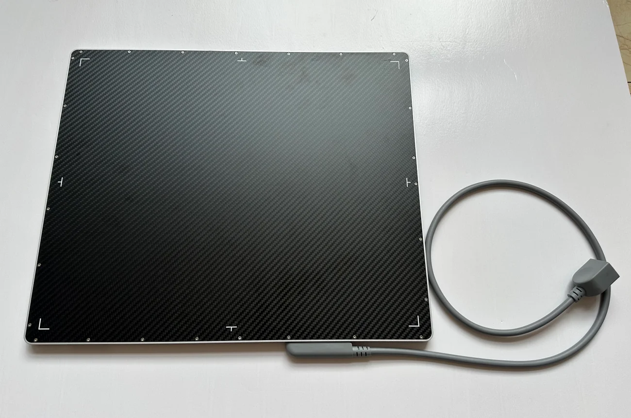
3072 x 3072 active pixels, 139 micrometer pixel pitch, 16-bit conversion and DQE of at least 70%.
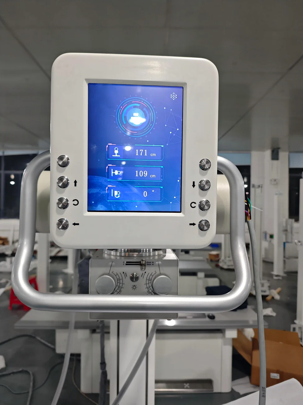
Examination parameters and motorized positioning controls are presented on a dedicated control panel.
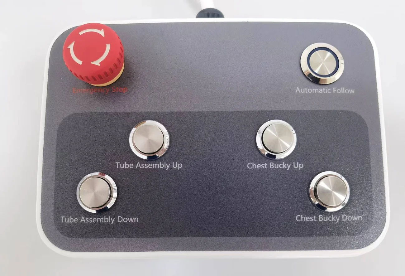
Dedicated controls support tube assembly and chest bucky movement during room positioning.
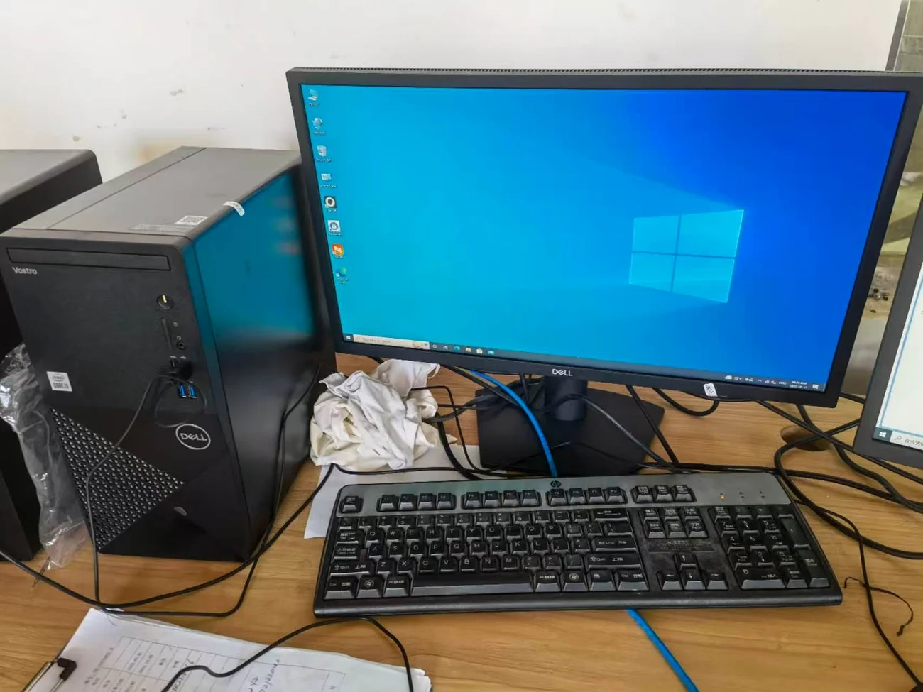
Multilingual acquisition, multiple APR positions, DICOM 3.0 and connection to RIS, PACS and printers.
Typical 50 kW configuration. Final component models and options are confirmed in the quotation.
| Model | YJ-FDR200-50 |
|---|---|
| System type | Floor-mounted digital radiography |
| Applications | Head, limbs, chest, spine, lumbar spine, orthopedics, emergency and operating-room workflows |
| Main control | 9.7-inch high-resolution touch screen |
| Table size | 2200 x 800 x 650 mm |
| Table load | 250 kg |
| Table travel | Front/back 120/120 mm; left/right 450/450 mm |
| Tube stand travel | Up/down 60-160 cm; left/right 50-180 cm |
| Tube rotation | -30 to 120 degrees; optional stand rotation -180 to 180 degrees |
| Bucky / FPD cover | 30-190 cm |
| Type | Amorphous silicon with CsI scintillator |
|---|---|
| Active area | 17 x 17 in (43 x 43 cm) |
| Active pixels | 3072 x 3072 |
| Pixel pitch | 139 micrometers; 100 micrometers optional |
| A/D conversion | 16 bit |
| DQE | At least 70% |
| Spatial resolution | 36 lp/cm |
| Acquisition time | 2 seconds or less |
| Detector option | Wireless detector optional |
| Generator output | 50 kW |
|---|---|
| Current | 10-630 mA |
| Voltage | 40-150 kV |
| mAs | 0.1-630 mAs |
| Exposure time | 0.001-6.3 s |
| Rated voltage | 380 VAC, plus or minus 10%, 50 Hz, 3-phase |
| Tube output power | 50 kW |
| Focal spot | 0.6 / 1.2 |
| Anode speed | 2800 rpm at 50 Hz / 3200 rpm at 60 Hz |
| Anode heat storage | 300 kHU |
| Maximum heat dissipation | 750 W |
| Collimator | Manual; maximum 450 x 450 mm window at 100 cm SID |
|---|---|
| Collimator light | LED, AC/DC 24 V, 5 W; 30-second timer; 1.5 mm Al inherent filtration |
| Workstation | 24-inch LCD; CPU 2.8 GHz or higher; memory 4 GB or higher; HDD 1 TB or higher |
| Network | Gbps network; Windows operating system |
| Software | Multiple languages, multiple APR positions, DICOM 3.0, RIS/PACS/printer connection |
| Grid option | 40 lp/cm; 10:1 ratio; 18 x 18 in; 100 cm table / 180 cm bucky focal distance |
| Ambient temperature | 10-40 degrees C |
| Relative humidity | 30-75% |
| Atmospheric pressure | 70-106 kPa |
This page reflects the supplied YJ-FDR200-50 product sheet. Final generator, tube, detector, grid, table, workstation, electrical supply, software and accessory configuration must be confirmed for each project and destination market.
The system is intended for head, limb, chest, spine and lumbar-spine radiography in orthopedics, emergency rooms, operating rooms and general imaging departments. Upright detector positioning also supports standing knee AP examinations.
We will review the intended use, electrical supply, image workflow and destination requirements before confirming the final configuration.