Compact mobile frame
A narrow wheeled base and folding tube arm support movement through wards, corridors and treatment rooms.
The YJ-M32DR combines a 32 kW X-ray generator, rotating-anode tube, wireless flat panel detector, folding tube arm and a 19-inch touchscreen workstation. It is configured for wards, emergency rooms, operating rooms, ICU and other bedside imaging environments.
The detailed specification sheet lists 40-125 kV, while the marketing sheet lists up to 150 kV. The final generator and tube rating must be confirmed by model and order before publication or quotation.
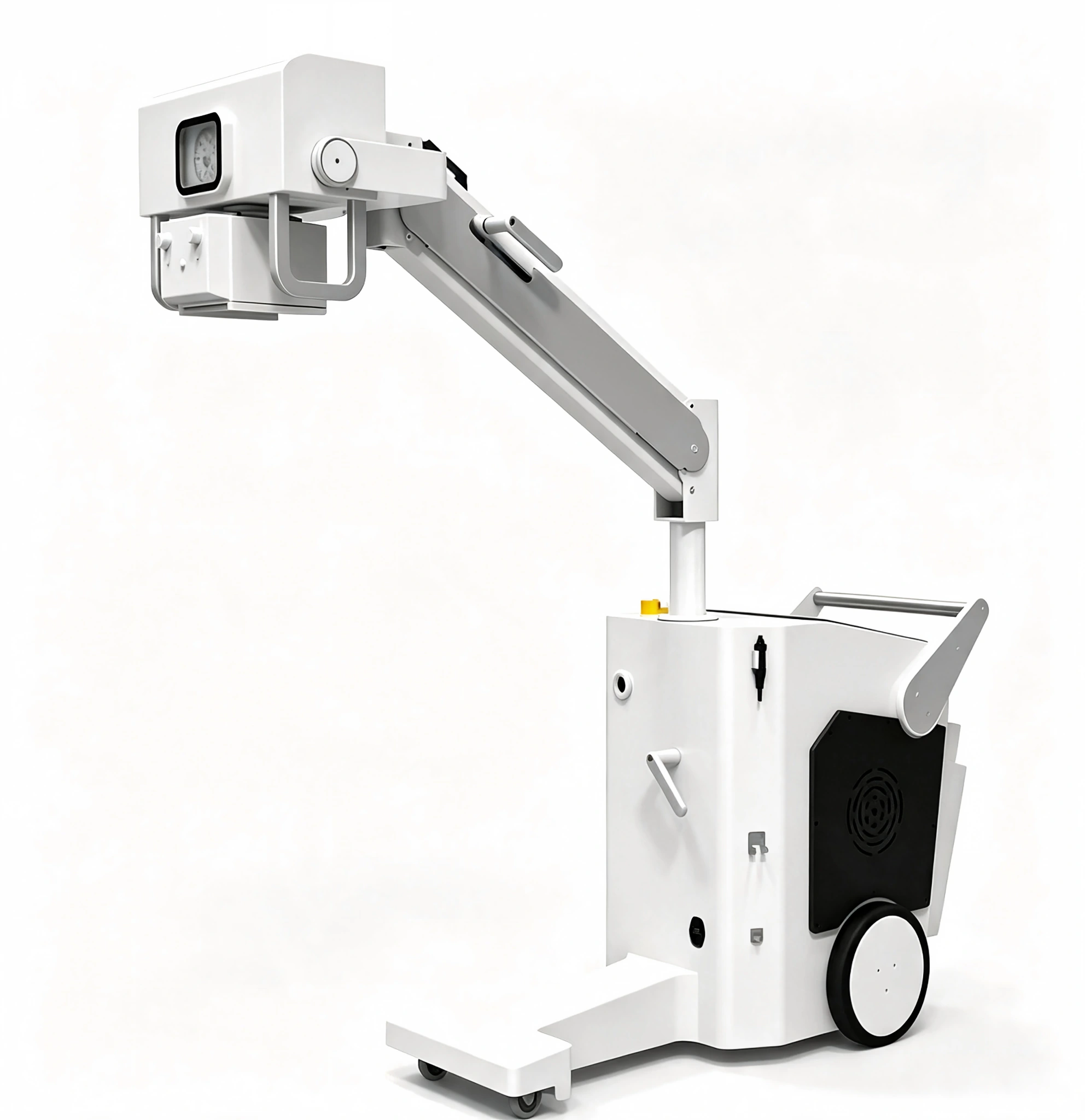
The white-background views show the complete mobile system. The factory image shows the assembled unit during preparation.
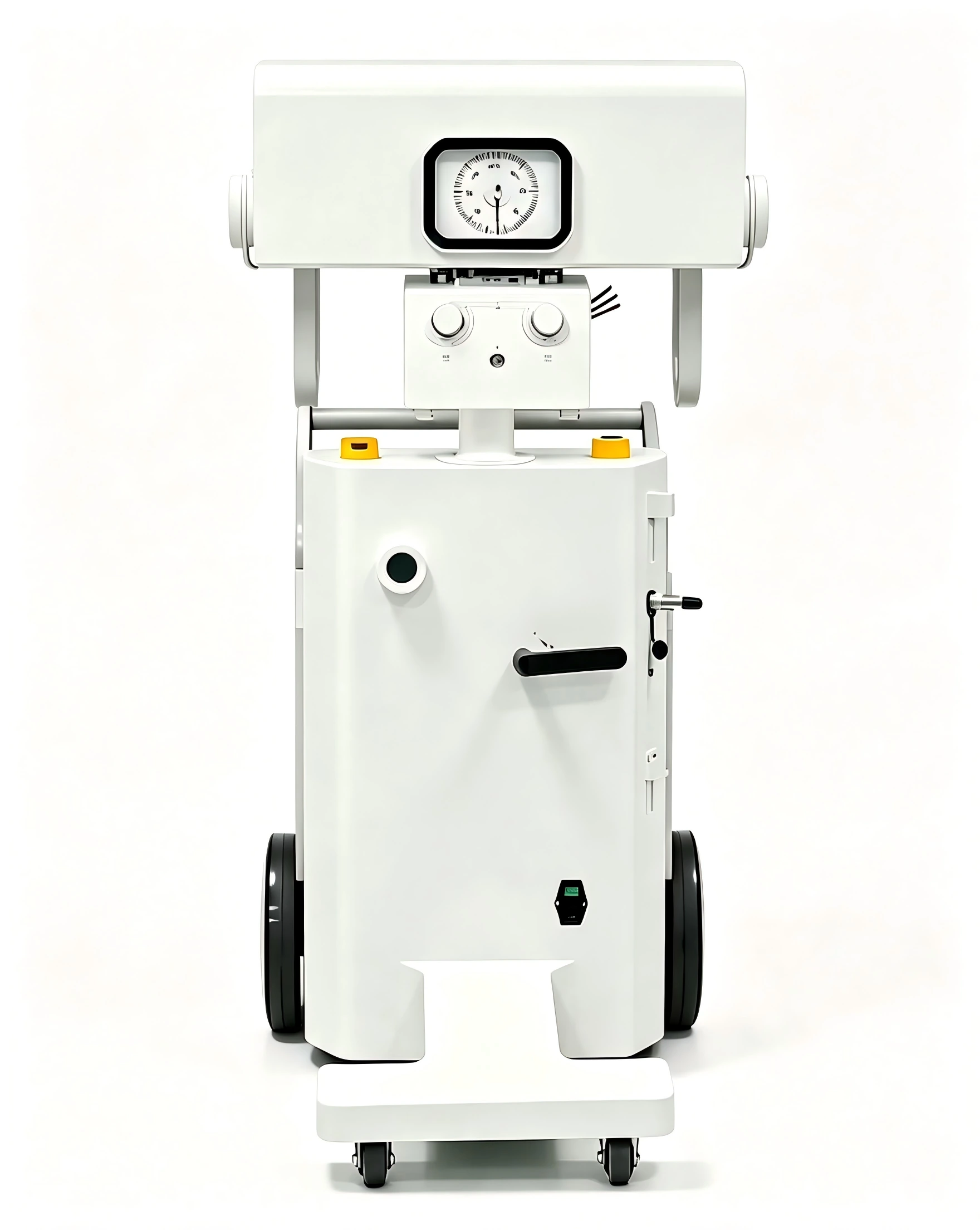
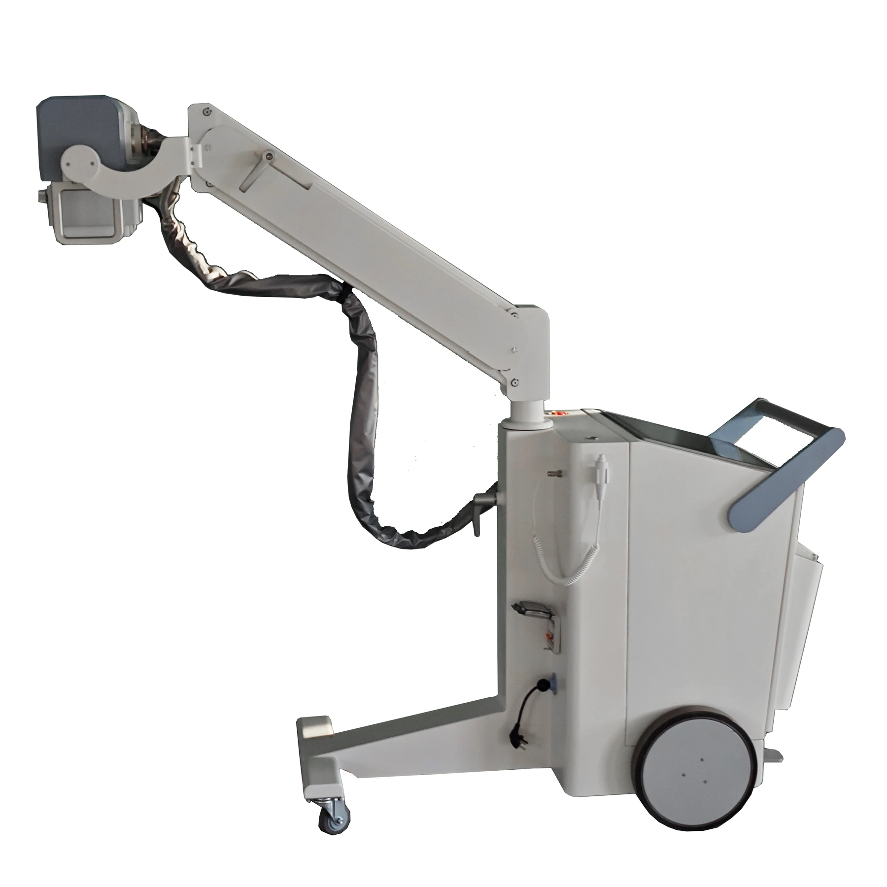
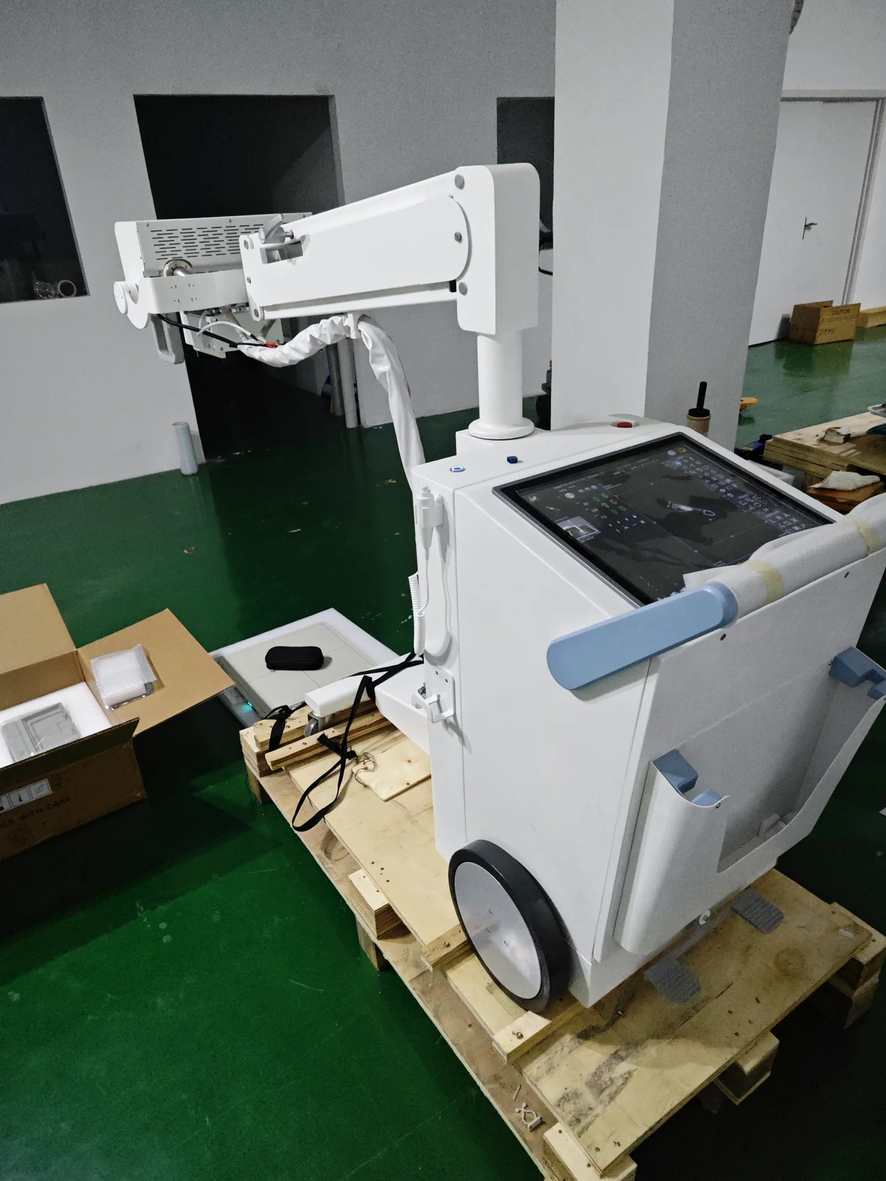
The supplied configuration focuses on mobility, simple positioning, wireless imaging and immediate image review.
A narrow wheeled base and folding tube arm support movement through wards, corridors and treatment rooms.
The wireless flat panel detector removes cable constraints and supports flexible positioning around the patient.
A 32 kW high-voltage generator and rotating-anode tube provide output for bedside radiography.
One-button power control, brake-step control, ultrasonic ranging and on-board image review support routine use.
Real configuration images and the supplied structure drawing show the main assembly, detector and workstation.
A rotating-anode tube and high-voltage generator are integrated into the mobile base.
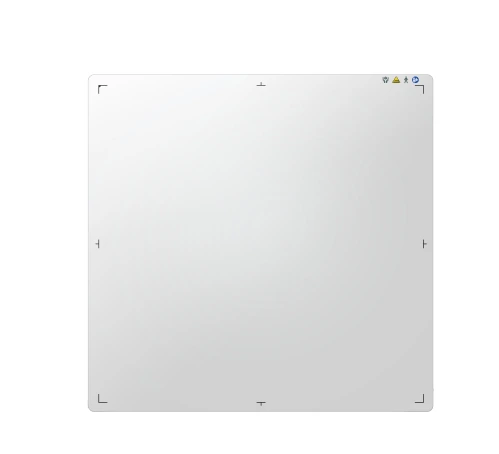
3072 x 3072 active pixels, 16-bit conversion, DQE of at least 70% and AED support.
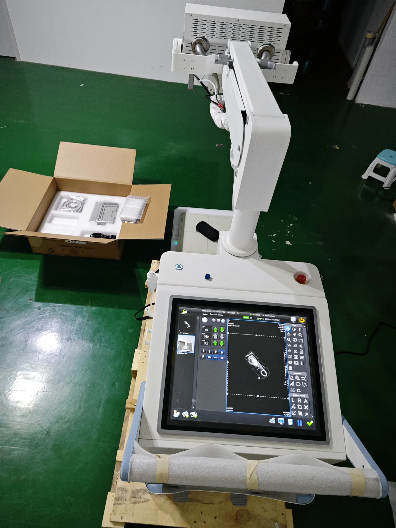
On-board image review, exposure setup, 1280 x 1024 display and DICOM 3.0 integration.
Tube focus distance from 490 to 2015 mm, with folding arm and vertical rotation for bedside exams.
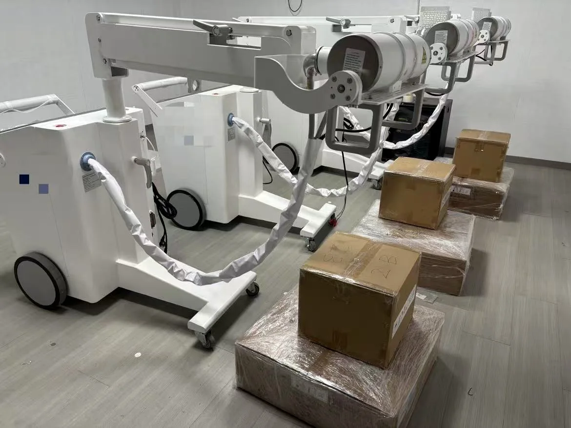
Multiple mobile DR systems shown during export preparation and pre-shipment handling.
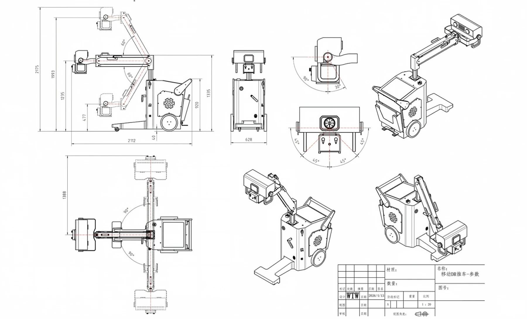
Supplied drawing supports discussion of the folding arm, tube rotation and system footprint.
Based on the detailed specification sheet. The voltage discrepancy is noted for final confirmation.
| Model | YJ-M32DR |
|---|---|
| Applications | Wards, emergency rooms, operating rooms, ICU and bedside radiography |
| Anatomy | Head, limbs, chest, waist, abdomen and other general radiography |
| Output power | 32 kW |
| Frequency | 25 kHz, plus or minus 10% |
| Current | 10-400 mA |
| Voltage | 40-125 kV in the detailed specification sheet; the marketing sheet lists up to 150 kV. Confirm final model. |
| mAs | 0.1-160 mAs |
| Exposure time | 1-6300 ms |
| Protection | Self-protection system with alarm and fault-code review |
| Detector type | Amorphous silicon wireless flat panel detector |
|---|---|
| Scintillator | Cesium iodide |
| Active area | 17 x 17 in (43 x 43 cm) |
| Available size | 14 x 17 in or 17 x 17 in, depending on configuration |
| Active pixels | 3072 x 3072 |
| Pixel pitch | 139 micrometers or less |
| A/D conversion | 16 bit |
| DQE | At least 70% |
| Spatial resolution | 35 lp/cm |
| Acquisition time | 2 seconds or less |
| AED | Equipped |
| Anode type | Rotating anode |
|---|---|
| Focal spot | 0.6 mm / 1.2 mm |
| Anode speed | 2800 rpm |
| Inherent filtration | 1.0 mm Al |
| Anode heat storage | 150 kJ (200 kHU) |
| Heat storage capacity | 900 kJ (1250 kHU) |
| Anode angle | 12.5 degrees |
| Maximum anode heat dissipation | 500 W |
| Tube support | Folding arm |
| Focus distance to ground | 490-2015 mm, plus or minus 5% |
| Arm folding range | -45 to +45 degrees |
| Tube tilt | Plus or minus 90 degrees |
| Vertical rotation | 360 degrees |
| Collimator axial rotation | Plus or minus 360 degrees |
| Collimator type | Manual collimator |
|---|---|
| Maximum window | 440 x 440 mm at 100 cm SID |
| Lamp | AC/DC 24 V, 5 W; LED illumination |
| Lamp timer | Automatic illumination with 30-second timer |
| Collimator filtration | 1.0 mm Al inherent filtration |
| Display | 19-inch LCD touch screen, maximum resolution 1280 x 1024 |
| CPU / memory | CPU 2.0 GHz or higher; memory 8 GB or higher |
| Storage | 512 GB or higher |
| Operating system | 64-bit Windows |
| DICOM | DICOM 3.0; PACS integration, patient data and image management |
| Environment | 10-40 degrees C; 30-75% relative humidity; 70-106 kPa |
The supplied documents contain a voltage-range difference and optional detector sizes. Confirm the exact generator, tube, detector, workstation, software and accessory configuration for each project.
The mobile system is intended for bedside and in-hospital radiography when moving the patient to a fixed room is impractical. The folding arm, wireless detector and on-board workstation support fast setup and image review.
We will review the workflow, detector size, generator configuration and market requirements before confirming the quotation.